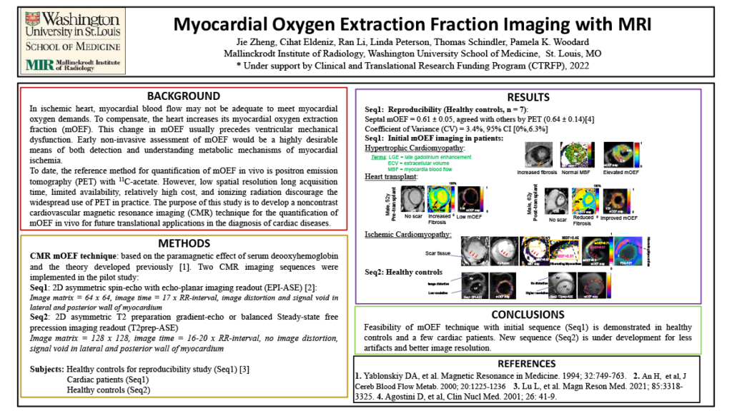
Introduction: Imbalance of myocardial oxygen supply and demand precipitates a cascade of physiological changes resulting in ischemic pathology. Myocardial oxygen extraction fraction (mOEF) may provide accurate assessment of this balance. Current non-invasive reference method to quantify mOEF is positron emission tomography (PET), but the low spatial resolution, high cost, and ionizing radiation discourage the widespread use of this method. Hence, the objective of this preliminary study is to develop and evaluate a new magnetic resonance imaging (MRI) technique for fast quantification of mOEF in vivo. Two specific aims will be addressed: 1) the reproducibility of this MRI method in vivo; 2) the diagnosis utility in patients with hibernating myocardium.
Methods: A new MRI acquisition method was recently developed for the quantification of mOEF with good image quality. Ten healthy adults will undergo cardiac MRI exams at two different days. Field inhomogeneity and motion correction will be two key components to address. We will then evaluate 10 patients with hibernating myocardial disease who are routinely diagnosed using 18F-FDG-PET for features of preserved glucose uptake (viable myocardium) and reduced blood flow. The mOEF will provide direct measurement of oxygen utilization, with hypothesis of increased mOEF for viable myocardium and reduced mOEF for non-viable myocardium.
Results: First, we expect our new imaging technique to allow consistent and reproducible good image quality and obtain correct global resting mOEF (coefficient of variation < 5%). Second, our hypothesis is confirmed in patients with hibernating myocardium.
Impact: This T1 work uses non-invasive and non-irradiation MRI to measure myocardial oxygenation for the fast diagnosis of oxygen deficiency in patients with cardiac ischemia. The finding could potentially guide therapy for functional recovery of myocardial contractility. The technique could improve health care quality by providing direct myocardial oxygenation assessment and monitoring treatment efficacy. Without using any exogenous tracers, this method can be combined with other cardiac MRI exams for “one-stop-shopping” test, thereby reducing healthcare costs.
Organization – Washington University in St. Louis
Zheng J, Eldeniz C, Schindler TH, Li R, Peterson LR, Woodard PM
