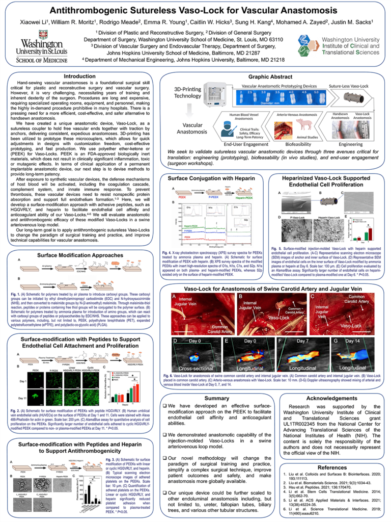
Introduction: Micro- and macrovascular anastomoses involve suturing together of blood vessels, which is a critical foundational surgical skill. However, it is costly, time-consuming, and prone to complications. Our team has created a unique anastomotic device, Vaso-Lock, as a sutureless coupler. Vaso-Lock is intraluminal, holding free vascular ends together with traction by anchors. In terms of its clinical application, we will devise methods to provide long-term patency. We aim to characterize a surface-functionalization method for Vaso-Locks to facilitate endothelial cell affinity and antithrombogenicity; and test anastomotic and antithrombogenic efficacy of Vaso-Locks in a swine carotid-jugular arteriovenous loop model.
Methods: We utilized 3D-printing to prototype Vaso-Locks with quick design adjustments. We apply plasma treatment to modify Vaso-Locks to conjugate peptides or polysaccharides. Endothelial cell and platelet adhesion assays will be implemented to evaluate biocompatibility and hemocompatibility. Angiography and Doppler ultrasound will be used longitudinally to assess patency, blood flow velocities, and lumen preservation in a swine model. Histological studies will be performed for endothelialization and device-vessel interface biocompatibility.
Results: Vaso-Locks can be deployed in porcine carotid arteries within 1 minute in comparison to handsewn anastomosis for 1 hour. The Vaso-Lock displayed higher tensile strength than that of handsewn anastomosis. Surface-modified with adhesive peptides encouraged greater endothelial cell attachment. Anastomosis was successfully achieved in the arterio-venous anastomosis. Doppler ultrasonography confirmed venous and arterial blood mixing throughout Vaso-Locks. Survival studies are underway to assess long-term patency.
Impact: This project will not only develop a new device for vascular anastomosis, but establish an effective surface modification approach for antithrombogenicity and endothelium formation. Our unique framework can potentially be applied to all luminal structures, which will simplify complex surgical techniques, improve patient outcomes and safety, and make anastomosis more globally available and feasible.
Organization – Washington University in St. Louis
Li X, Moritz W, Meade R, Kang S, Zayed M, Sacks J
